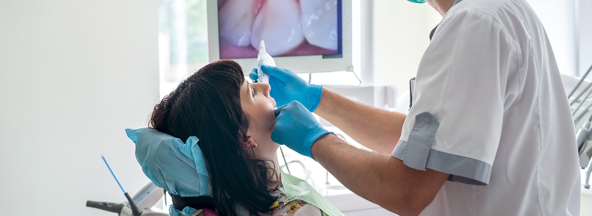
An intraoral camera is a compact, pen-sized imaging device that captures high-resolution, full-color pictures from inside the mouth. Unlike traditional mirrors, this tool delivers a magnified, real-time view of teeth, gum tissue, and other oral structures directly to a monitor. The result is a level of visual detail that supports more accurate observation of surface defects, restorative margins, and soft tissue conditions without requiring the patient to visualize awkward angles themselves.
Technically simple to use but clinically powerful, the intraoral camera typically employs LED lighting and a small sensor to produce clear images even in tight, hard-to-see spaces. Captured frames can be paused, zoomed, and reviewed on-screen for immediate discussion. That capability helps clinicians explain findings with visual evidence, so patients can follow along and better understand the nature and extent of any concerns.
Because these images are digital, they integrate smoothly into modern dental workflows and electronic records. The immediacy of the visual information shortens the gap between examination and explanation, enabling more informed conversations during the same appointment. For patients who prefer to see what the dentist sees, the intraoral camera turns abstract descriptions into concrete images.
One of the most practical benefits of intraoral imaging is how it enhances communication. When clinicians show patients a close-up photo of a fractured filling, an early cavity, or inflamed gum tissue, the diagnosis becomes easier to understand. This shared visual reference encourages questions and helps patients make decisions based on a clearer grasp of their oral health rather than relying solely on verbal descriptions.
Images from an intraoral camera also serve as a teaching tool during routine exams. Patients can see plaque accumulation, wear patterns, or enamel erosion that might otherwise go unnoticed. That transparency often increases patient engagement with preventive steps—such as improved brushing technique or targeted home care—because the need for change is visible and specific.
Clinicians can annotate or magnify particular areas on-screen to emphasize problem zones and explain recommended next steps. This collaborative approach builds trust and reduces anxiety, especially for patients who feel more secure when they understand the evidence behind a recommendation. In short, intraoral cameras help turn clinical observations into a two-way conversation.
Intraoral cameras are versatile diagnostic assistants across a broad spectrum of routine and advanced care. They help detect early caries, hairline cracks, failing margins on restorations, and soft tissue changes that warrant closer attention. Early detection is a major advantage because small problems are often easier to treat and manage than those discovered at later stages.
Beyond diagnosis, these cameras are valuable for monitoring progress over time. Sequential images taken across appointments let clinicians track healing after periodontal therapy, the integrity of restorations, or the evolution of enamel defects. This chronological visual record supports evidence-based decisions and allows adjustments to care plans based on observable trends rather than memory alone.
In specialty care, intraoral images can assist in treatment planning for restorative work, prosthodontics, and implant-related procedures by providing detailed views of occlusal anatomy and soft tissue contours. While not a substitute for three-dimensional imaging where bone assessment is required, intraoral cameras complement other diagnostic tools by emphasizing surface-level detail and soft-tissue health.
When an image is captured, it becomes part of the patient’s permanent digital record, where it can be referenced in future visits or included when consulting with colleagues. These archived images help create a consistent, visual narrative of a patient’s oral health, which can be essential when coordinating care with specialists, dental laboratories, or other members of a treatment team.
Digital files are easy to export or attach to referral documentation, improving the clarity and efficiency of collaborative case discussions. For instance, a clear intraoral photograph can help a periodontist or prosthodontist understand the clinical situation before a consultation, reducing the need for redundant examinations and speeding up decision-making.
Within our office technology ecosystem, intraoral photos work alongside digital x-rays, CBCT scans, and CAD/CAM data to create a fuller picture of oral health. That cohesive approach supports comprehensive treatment planning and helps ensure that clinical choices are grounded in multiple, corroborating forms of evidence.
An intraoral camera exam is quick, noninvasive, and fully integrated into a standard dental visit. The clinician or assistant gently maneuvers the camera inside the mouth while the patient relaxes in the chair. Most patients experience no discomfort beyond the normal sensation of having an instrument near the teeth, and the live images are displayed on a monitor for immediate review.
During the examination, the clinician will typically capture several images of targeted areas—such as questionable restorations, tight contacts, or suspicious soft tissue sites. Each photo is reviewed on-screen and saved to the patient’s chart when needed. The process adds only a few minutes to a routine exam but delivers a significant amount of diagnostic and educational value.
For patients concerned about radiation or invasive testing, intraoral cameras offer a radiation-free way to evaluate surface conditions. They are an especially helpful option for those who appreciate visual explanations of their care. If follow-up imaging or additional diagnostics are recommended, the camera images provide a clear baseline that can guide next steps.
Wrap-up: Intraoral cameras bring clarity, documentation, and better communication to modern dental care. They help clinicians detect subtle problems earlier, involve patients more fully in their treatment, and create a visual record that supports coordinated care. To learn how intraoral imaging fits into your dental visits, contact us for more information.
An intraoral camera is a pen-sized, high-resolution camera designed to photograph the interior of the mouth and display live images on a monitor. It provides full-color, close-up views of teeth, restorations, and surrounding soft tissues to aid in examination and documentation. The device captures both still images and short video clips that can be saved for review and comparison over time.
Because the camera is small and maneuverable, it can access areas that are difficult to see with the naked eye or standard mirrors. Real-time imaging helps clinicians identify early signs of decay, fractures, wear, and soft tissue changes. Saved images become part of the patient record and support clear communication about diagnosis and treatment planning.
An intraoral camera enhances diagnostic accuracy by providing magnified, well-lit images that reveal details that may be missed during a visual exam. High-resolution images help clinicians detect small cracks, recurrent decay around restorations, and subtle soft tissue abnormalities. The enlarged view reduces uncertainty and supports more precise clinical decision making.
Images can be reviewed frame by frame or compared with prior records to monitor progression or healing. This visual documentation assists in prioritizing care and selecting minimally invasive treatment options when appropriate. Together with other diagnostic tools, intraoral imaging contributes to earlier detection and better outcomes.
Images taken with an intraoral camera are stored digitally as part of the patient's chart and are referenced during follow-up visits and treatment planning. These files create a visual timeline that documents the condition of teeth and soft tissues over months and years. Digital storage allows for easy retrieval and secure long-term record keeping.
Captured images also streamline communication with other members of the dental team, dental laboratories, and specialists when collaborative care is needed. Having clear, dated images reduces ambiguity and supports continuity of care across providers. This documentation can also assist with insurance documentation when a clinical image is required to substantiate treatment recommendations.
Yes, patients can view intraoral camera images in real time on a monitor, which helps them understand the clinician's observations and recommended treatments. Seeing detailed photos of their own teeth and gums often clarifies clinical findings and allows patients to make more informed decisions. Visual information enhances the education process by linking spoken explanations to concrete images.
When patients can examine images alongside the clinician, it encourages questions and shared decision making. This transparency builds trust and helps align treatment goals with the patient's expectations. Viewing images also makes it easier to track improvements after restorative or periodontal therapies.
Intraoral camera imaging is noninvasive and generally very comfortable for patients, as the device is small and handheld with no radiation exposure. The procedure typically involves brief positioning of the camera inside the mouth to capture targeted views, and most patients experience little or no discomfort. Because it is purely optical, the camera does not introduce any clinical risk beyond routine oral examination.
Clinicians follow standard infection control protocols, including barriers and sterilization procedures for any components that contact the patient, to ensure safety. The short duration of imaging and the minimal physical contact contribute to a positive patient experience. If a patient has sensitivity or a strong gag reflex, the clinician can modify technique to enhance comfort.
High-quality intraoral images provide laboratories and specialists with precise visual information about tooth shape, color, margin position, and soft tissue contours. Sharing these images helps technicians fabricate crowns, veneers, and appliances that better match the clinical situation. Images can be sent electronically to support remote consultation and coordinated treatment planning.
When a specialist receives clear photographs, they can more quickly assess referral needs and recommend targeted interventions. Visual records reduce the need for multiple in-person evaluations and help ensure that laboratory work aligns with the clinician's expectations. This efficiency supports smoother workflows and improved clinical outcomes.
Intraoral camera images complement other digital tools such as CBCT, digital impressions, and chairside CAD/CAM systems by adding detailed surface-level visuals to three-dimensional or scanned data. These combined data sets give clinicians a more complete view of the clinical situation, enabling more accurate treatment planning and execution. For example, camera photos can be paired with digital scans to refine shade selection and restoration margins.
Integration with practice management software and imaging platforms allows clinicians to view and compare multiple modalities within a single interface. This interoperability streamlines workflows and enhances the precision of procedures like same-day restorations and guided implant placement. The combined digital approach supports a higher level of diagnostic confidence and treatment predictability.
Cosmetic Micro Dentistry uses intraoral camera images to document baseline conditions, confirm clinical findings, and communicate proposed treatment steps with patients. Real-time photos allow the team to point out areas of concern, demonstrate how different treatments will address specific issues, and discuss alternatives in a visual, patient-friendly way. This approach supports collaborative treatment planning and informed consent.
Images are also referenced when coordinating care with specialists or when fabricating restorations in-house with the Glidewell Fastmill. Visual documentation helps ensure restorative designs and surgical guides reflect the patient's actual anatomy and aesthetic goals. The result is a more predictable and efficient treatment experience.
During an intraoral camera exam, the clinician will position the small camera inside the mouth and capture a series of targeted images while the patient remains seated comfortably. The process is quick and typically takes only a few minutes, with images displayed on a monitor so the patient can view them as they are taken. There is no radiation and no invasive steps associated with this imaging method.
Patients may be asked to open wide, move their tongue, or change head position briefly to obtain the best views. If necessary, the clinician will explain each image and answer questions about what the pictures reveal. The images are then saved to the patient record for future reference and comparison.
Clinics follow strict infection control protocols to clean and disinfect intraoral camera housings and to protect any parts that contact the oral mucosa. Disposable barrier sleeves are commonly used for the camera head during each patient encounter, and reusable components are cleaned and sterilized according to manufacturer guidelines. These measures reduce cross-contamination risk and align with standard dental infection control practices.
Staff receive training in proper handling and reprocessing of camera components to ensure consistent compliance with safety standards. Documentation of cleaning procedures and adherence to published protocols help maintain a safe environment for patients and clinical teams. If a patient has specific concerns about infection control, the team can review the steps taken during their appointment.
Quick Links
Contact Us