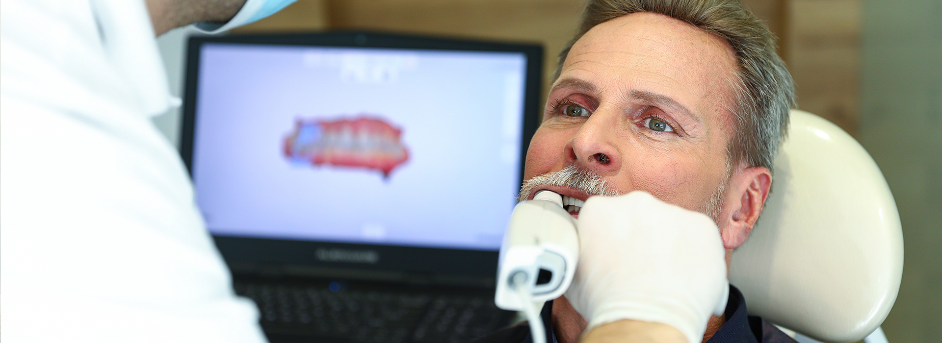
Digital impressions use advanced intraoral scanners to create a precise three-dimensional record of a patient’s teeth and surrounding soft tissues. Instead of taking a traditional putty impression, the clinician moves a small wand—equipped with cameras and light sources—around the dental arches. The wand captures a series of images and depth data that software stitches together into a continuous, accurate 3D model.
These digital models are more than pictures; they are volumetric files that show exact tooth contours, interproximal contacts, and the subtle transitions between tooth and gum. The same data can be used for treatment planning, laboratory communication, and direct CAD/CAM fabrication. Because the scan is immediate and visible on-screen, clinicians can evaluate margins, occlusion, and soft-tissue relationships in real time and re-scan any missing detail on the spot.
Adopting digital impressions changes how practices operate at a fundamental level. The technology reduces many manual steps associated with plaster models and physical shipping, and it creates a durable, searchable digital record that supports long-term case management. For practices focused on predictable, efficient care, digital impressions are an essential upgrade.
One of the most noticeable benefits of digital impressions is the improvement in patient comfort. Traditional impressions require trays filled with viscous material that can trigger gag reflexes and cause discomfort for many patients. Scanning eliminates this need—there’s no tray, no material setting against the palate, and no unpleasant taste or texture to tolerate.
Beyond comfort, digital scanning speeds up chair time and reduces back-and-forth appointments. Immediate visualization empowers patients to see what the dentist sees, which helps communication and informed decision-making. For anxious patients or those with sensitive gag reflexes, the calmer, quicker scanning process often makes treatment planning and restorative appointments easier to accept.
An additional practical advantage is improved infection control. Scanning wands are designed for cleaning and disinfection, and the elimination of impression materials reduces handling of biological matter that would otherwise be poured into stone models. These features contribute to a safer, more streamlined patient pathway from diagnosis to restoration.
Accuracy is at the core of why digital impressions are preferred for many restorative workflows. High-resolution scanners can capture fine details of preparation margins and adjacent teeth, producing files that support tight-fitting crowns, inlays, and bridges. When margins are clear and data capture is complete, laboratory or in-office milling systems can reproduce restorations with fewer adjustments.
Clinical outcomes improve because the digital workflow reduces distortion and variability. Traditional impressions can distort during removal, transport, or when poured into stone; digital files remain consistent from capture to fabrication. This consistency often means fewer remakes and less chairside adjustment because restorations arrive closer to the intended shape and contact scheme.
Beyond dimensional accuracy, software tools help clinicians refine scans by identifying undercuts, verifying margin clarity, and simulating occlusion. This diagnostic layer allows proactive correction before sending a case for fabrication, elevating the predictability of both simple and complex restorative procedures.
Digital impressions are the entry point to a fully digital restorative workflow. After a scan is captured, the data can be used immediately for virtual design (CAD), sent electronically to a trusted dental laboratory, or routed to an in-office milling unit for same-day restorations. This flexibility supports a wide range of treatment timelines—from multi-appointment cases to single-visit crowns and onlays.
When a practice integrates scanning with in-office CAD/CAM milling, patients can often receive permanent ceramic restorations in one appointment. The scan supplies the 3D model, design software allows precise customization of form and occlusion, and the milling system fabricates the restoration from a solid ceramic block. The result is a seamless, end-to-end process that reduces interim steps and shortens overall treatment time.
Even when a laboratory is involved, electronic transmission simplifies collaboration. Digital files travel instantly, enabling lab technicians to begin work sooner and review scans with the clinician if questions arise. This rapid exchange improves coordination and helps maintain clinical momentum while delivering high-quality outcomes.
Finally, the digital workflow supports future care: archived scans serve as baseline records for monitoring wear, planning replacements, or guiding orthodontic and implant therapy. That longitudinal value makes digital impressions an investment in both immediate restorations and future diagnostic capability.
Modern intraoral scanners combine optical imaging with structured light or laser technology to capture reliable data quickly. Many units also record color and texture, which helps laboratories match shade and surface characteristics. These technical capabilities improve communication across the team and yield restorations that look and function more naturally.
Data security and storage are essential considerations in a digital practice. Scanned files should be managed through secure systems that comply with privacy standards and protect patient information. Practices typically use encrypted transmission when sharing files with external labs and maintain secure backups for internal archives. These measures safeguard both patient privacy and the integrity of clinical records.
Routine maintenance and staff training are important to keep scanning systems performing at their best. Regular calibration, software updates, and proper disinfection protocols ensure consistent image quality and reduce the risk of scanning artifacts. With an emphasis on upkeep and technique, teams can maximize the reliability of their digital impression workflows.
Incorporating digital impressions into daily practice requires thoughtful planning—but the clinical and operational benefits are substantial. Scanning enhances diagnostic precision, improves patient experience, and streamlines restorative workflows. Whether used for single crowns, multi-unit bridges, or implant planning, digital impressions support more predictable, efficient care pathways.
Clinicians and staff who embrace scanning often find that it reshapes how they approach case planning, lab communication, and patient education. The visual nature of scans helps patients understand proposed treatments, which in turn fosters clearer expectations and smoother treatment acceptance.
For practices committed to modern, high-quality dentistry, digital impressions are a cornerstone technology. Cosmetic Micro Dentistry integrates these tools into a broader digital ecosystem—supporting same-day restorations, improved clinical accuracy, and an elevated patient experience without compromising safety or record integrity.
Digital impressions replace messy trays with precise, three-dimensional data capture that benefits clinicians and patients alike. They improve comfort, enhance accuracy, and enable faster, more coordinated restorative workflows. With secure recordkeeping and routine system care, scanning becomes a durable asset for both immediate treatment and long-term case management.
If you’d like to learn more about how digital impressions are used in everyday restorative dentistry or how this technology might apply to your care, please contact us for more information.
A digital impression is a precise, computer-generated 3D model of your teeth and surrounding oral tissues captured with an intraoral optical scanner. Instead of using trays and impression paste, the clinician moves a small wand across the teeth to collect thousands of optical images that are stitched together into a continuous digital file. The result is an accurate replica that can be used for restorations, orthodontic appliances and diagnostic records.
Digital impressions eliminate the need to pour stone models or ship physical impressions to a laboratory, because the digital file can be transmitted electronically. They also enable clinicians to evaluate margins, contacts and occlusion on-screen and to make adjustments in real time. This workflow supports faster communication with dental labs and more predictable restorative outcomes.
An intraoral scanner uses a handheld imaging wand that emits structured light or laser patterns to capture detailed surface data from teeth and soft tissues. The device records multiple overlapping images which proprietary software aligns and fuses into a single three-dimensional model. During the scan the system registers color and texture information that helps clinicians and technicians assess shade, anatomy and soft-tissue relationships.
After capture, the software refines the mesh, removes artifacts and can register occlusion by scanning the bite. The final digital file is exportable in common formats for CAD/CAM design, laboratory collaboration or integration with other technologies such as CBCT. This digital interoperability makes intraoral scans a versatile tool in modern restorative and implant workflows.
Digital impressions are typically more comfortable for patients because they eliminate bulky trays and impression material that can cause gagging or discomfort. They reduce the likelihood of retakes since clinicians can review the scan immediately and capture any missed areas in real time. The digital workflow also minimizes errors associated with material distortion, stone model inaccuracies and shipping damage.
For clinicians and labs, digital files speed up communication and support streamlined fabrication processes, including CAD/CAM design and in-office milling. Patients benefit from shorter treatment timelines and clearer visual explanations of their dental needs. Overall, the technology improves efficiency, predictability and patient experience without compromising clinical quality.
Modern intraoral scanners produce highly accurate digital impressions that are suitable for crowns, bridges and many types of implant restorations. Scanners capture fine details such as preparation margins and interproximal contacts, and laboratory systems can use these digital files to design restorations with precise fits. Clinical protocols often include verification steps to ensure adequate margin capture and occlusal relationships before fabrication.
When planning implant restorations, digital impressions can be combined with CBCT data to create comprehensive surgical and prosthetic plans. Guided implant systems and digital implant libraries accept these files to support accurate component selection and prosthesis fabrication. Proper scanning technique and clinician oversight remain important to achieve the best restorative fit and long-term outcomes.
Digital impressions eliminate physical shipping delays by allowing immediate electronic transfer of files to the dental laboratory or in-office milling system. This rapid transmission reduces back-and-forth waits and often shortens the time from preparation to final restoration. Because labs receive high-resolution digital data, they can design and mill restorations more efficiently and with fewer manual adjustments.
In practices equipped with in-office CAD/CAM milling, digital impressions can be used to produce same-day restorations, removing the need for temporary crowns and multiple visits. Even when external labs are involved, digital workflows typically result in fewer remakes and faster turnarounds due to improved consistency and clearer communication. The overall result is a faster, more streamlined patient experience.
Yes. Digital impressions are generally more comfortable than traditional impressions because they do not require filling the mouth with impression material or holding large trays in place. The scanning process is quick and noninvasive, and most patients tolerate it well, including those with sensitive gag reflexes. Intraoral scanners rely on optical imaging rather than ionizing radiation, so the procedure itself does not expose patients to additional radiation.
Clinicians will evaluate each patient’s ability to tolerate an intraoral scan and use techniques such as retraction or sectional scanning to capture difficult areas. Pediatric patients and patients with special needs often find digital scanning less stressful than conventional methods. Safety protocols for handling, storing and transmitting digital records further protect patient information and clinical integrity.
Digital impressions are a core component of same-day restorative workflows because they provide immediate, accurate digital data that can be sent directly to CAD/CAM software for design. Once the restoration is designed, it can be milled in-office from ceramic or composite blocks and then stained, glazed and seated during the same appointment. This integrated process removes the need for temporaries and multiple fitting visits when clinical conditions allow.
Many practices use an in-office milling unit and a validated restorative material to complete crowns, onlays and certain implant prostheses in a single visit. Cosmetic Micro Dentistry employs these digital workflows to increase efficiency and patient convenience while maintaining clinical standards for fit and aesthetics. Proper case selection and laboratory collaboration remain important to ensure predictable results.
Digital impression data is treated as protected health information and is managed according to applicable privacy and security standards. Files are typically stored on secure office servers or cloud systems with encryption during transmission to labs or production centers. Access controls, user authentication and audit trails help ensure that only authorized team members and partners can view or modify the data.
Dental practices establish retention and backup policies to preserve clinical records and to support continuity of care. When files are shared with external laboratories or specialists, secure file-transfer protocols and contractual privacy safeguards are used to protect patient information. Patients may request access to their digital records through standard office procedures.
Yes. Digital impressions enhance treatment planning by providing detailed, manipulable 3D models that improve visualization of anatomy, occlusion and soft-tissue relationships. Clinicians can simulate restorative designs, evaluate esthetics and verify functional contacts on-screen before any restoration is fabricated. This digital preview supports informed decision-making and helps patients understand proposed treatments.
Digital files also integrate with other technologies such as CBCT imaging and guided surgery systems to create comprehensive interdisciplinary plans, particularly for implant cases. The ability to collaborate with laboratories and specialists using shared digital data fosters more predictable surgical guides and prosthetic outcomes. Overall, digital impressions make planning more precise and collaborative.
During a digital impression appointment the clinician will first explain the process and position you comfortably in the dental chair. A small wandlike scanner is then moved around your teeth and gums to capture a series of images that the system assembles into a 3D model; the scanning portion usually takes only a few minutes depending on the scope of the work. You may be asked to bite together briefly for a bite registration and to rinse or allow the clinician to retract soft tissues for clearer capture.
After the scan, the dentist will review the model on-screen with you to confirm margins, contacts and esthetic considerations and to discuss the next steps. If the practice offers in-office milling, the restoration may be designed and fabricated that day, or the file will be sent securely to a laboratory for final fabrication. At Cosmetic Micro Dentistry patients are shown their scans and given a clear explanation of how the digital data informs treatment sequencing and expected outcomes.
Quick Links
Contact Us