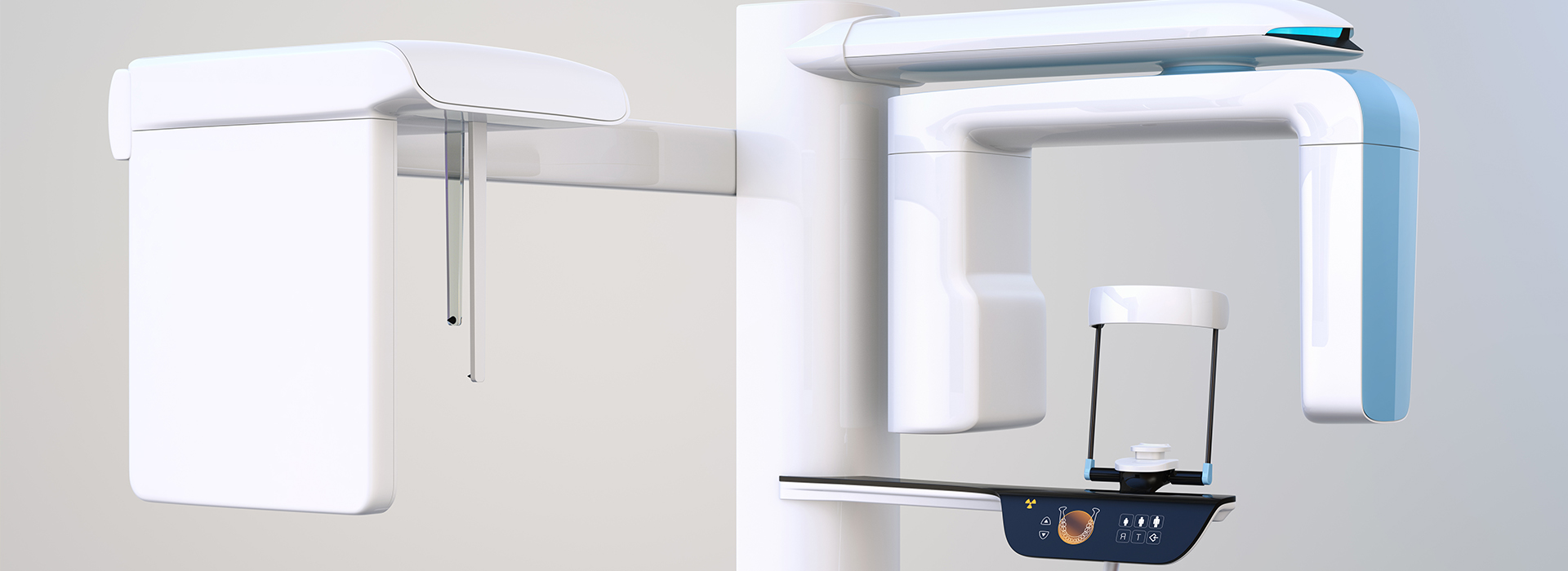
At the office of Cosmetic Micro Dentistry, we use the latest technology to provide our patients with the most precise and gentle care. We firmly believe that achieving successful treatment outcomes relies on using the most advanced diagnostic technology. By obtaining high-resolution and detailed 3D images, we can develop the right treatment plan and provide the highest level of care.
As a state-of-the-art practice, we utilize one of the most advanced CBCT (cone-beam computed tomography) imaging systems to obtain the sharpest 3D diagnostic films while exposing the patient to the lowest level of radiation.
With the advanced and versatile capabilities provided by our CBCT imaging system, it’s quick, safe, and comfortable to obtain detailed 3D views of a patient's teeth, jaws, and surrounding anatomy.
Because they obtain targeted, detailed, and distortion-free views from multiple angles, CBCT scans provide essential information for numerous analyses and the assessment of maxillofacial disorders or pathology. With applications in every area of dental care, CBCT imaging facilitates precise surgical planning, including the accurate placement of dental implants.
We value your time as well as your investment in oral health. Our office maintains an unwavering commitment to excellence and strives to provide the most advanced and comfortable solutions to meet your family’s dental needs. With patient care and comfort as our top priorities, you can depend on our practice for the highest quality of skilled and compassionate care.
Elevating diagnostic capabilities to an entirely new level, our practice is equipped with the 3D Accuitomo 170 by Morita, a leading-edge Cone Beam Computed Tomography (CBCT) unit. Unlike traditional 2D X-rays that provide a flat image, this advanced technology generates a precise 3D model of your jaw, teeth, and surrounding structures. This allows us to visualize intricate details like individual root canals, nerve pathways, and bone density with incredible clarity. With a focus on patient safety, the 3D Accuitomo 170 uses a minimal radiation dose, often significantly less than conventional CT scans or even a full mouth series of 2D X-rays. This high-resolution, low-dose imaging is an invaluable tool for precise treatment planning for implants, endodontics, orthodontics, and a variety of oral surgeries, ensuring a more accurate diagnosis and a more predictable outcome for your care.
CBCT stands for cone-beam computed tomography, a 3-D imaging method that captures volumetric data of the teeth, jaws and surrounding anatomy. Unlike traditional two-dimensional X-rays, CBCT produces cross-sectional and three-dimensional views that reveal depth, spatial relationships and internal structures that a flat film cannot show. This extra dimensional detail helps clinicians evaluate complex anatomy with greater confidence.
CBCT imaging collects multiple images in a single rotation and reconstructs them into a digital volume, which clinicians can view in slices or render as a 3-D model. This capability enables more accurate measurements of bone height, width and density, identification of impacted teeth and clear visualization of nerve pathways. In short, CBCT supplements conventional radiography when advanced diagnostic information is required for planning or problem solving.
CBCT scans are useful for a wide range of clinical applications, including evaluation of impacted or supernumerary teeth, assessment of jaw pathology, and visualization of root canal anatomy. They can reveal fractures, resorptive defects, cysts or tumors that may be difficult to detect on 2-D films, and they provide precise localization of critical structures such as the inferior alveolar nerve. This makes CBCT valuable for both routine and complex diagnostic challenges.
Because the images are distortion-free and can be viewed from any angle, clinicians use CBCT to assess sinus health prior to sinus-lift procedures, to evaluate temporomandibular joint (TMJ) anatomy, and to analyze airway space for sleep-related breathing concerns. The modality supports evidence-based decision-making across endodontics, oral surgery, orthodontics and implant dentistry by revealing information that influences treatment selection and sequencing.
At Cosmetic Micro Dentistry a typical CBCT appointment is brief and noninvasive: the patient remains seated or standing while the scanner rotates around the head to capture the image volume. The technologist will ask the patient to remain still and may provide a bite block or head support to reduce motion; metallic objects such as jewelry or eyeglasses should be removed before imaging. The scan itself usually takes less than a minute, and the total appointment time is commonly under 15 minutes including setup.
CBCT is generally comfortable and does not require any injections or nausea-inducing preparations, and most patients experience no discomfort during the capture. Once images are acquired they are reconstructed into a DICOM volume that the clinician reviews on dedicated software, allowing immediate assessment and discussion of findings with the patient. If additional interpretation is needed, images can be securely shared with specialists or an oral and maxillofacial radiologist for consultation.
CBCT uses ionizing radiation, so imaging decisions follow the principle of ALARA (as low as reasonably achievable) to balance diagnostic benefit and exposure. Modern CBCT units and targeted fields of view limit radiation by capturing only the area needed for diagnosis, and many dental CBCT scans deliver substantially less radiation than medical CT scans. When compared to a full-mouth series of conventional 2-D X-rays, a small-field CBCT exam can produce similar or only slightly higher doses while offering far greater diagnostic information.
Clinicians justify CBCT examinations based on individual patient needs and existing diagnostic data, avoiding routine or unnecessary use. Special precautions are taken for children and pregnant patients; imaging during pregnancy is generally avoided unless the information is essential for urgent care and cannot be obtained by other means. Your clinician will discuss risks and benefits and choose the smallest field of view and appropriate exposure settings for your situation.
CBCT provides the accurate three-dimensional measurements and spatial relationships necessary for predictable implant planning, including assessment of bone volume, bone quality and proximity to critical anatomy such as nerves and sinuses. These images allow the clinician to virtually place implants within the available bone and evaluate angulation, depth and restorative space prior to surgery. The result is a more precise surgical plan that reduces intraoperative guesswork and the risk of complications.
When used with digital planning software, CBCT data can be used to fabricate surgical guides or to drive dynamic navigation systems that translate the virtual plan to the clinical setting. This integration enhances accuracy in implant placement and can shorten surgical time and improve prosthetic outcomes. Careful preoperative imaging and planning are central to achieving long-term implant stability and optimal esthetic results.
Yes. CBCT is particularly helpful in endodontics for identifying complex root canal anatomy, locating accessory canals, and detecting vertical root fractures or persistent periapical pathology that may not be visible on 2-D radiographs. The modality can reveal the true extent and shape of lesions, helping clinicians determine whether nonsurgical retreatment or surgical intervention is indicated. In cases of ambiguous symptoms or failed prior therapy, CBCT often clarifies the diagnosis and guides targeted treatment.
CBCT also aids in planning apical surgery by mapping the location of roots relative to anatomic structures and showing lesion margins with precision. Because endodontic decisions can hinge on subtle anatomic detail, three-dimensional imaging frequently changes or refines treatment plans to improve the likelihood of a successful outcome. Interpretation should be performed by a clinician experienced in reading dental CBCT volumes.
Preparation for a CBCT scan is minimal: patients should remove removable metal objects such as earrings, necklaces and removable dental appliances that could create artifacts in the image. There is no need to fast or alter medications for routine scans, and the technologist will provide instructions to stabilize the head and minimize movement. Children may require extra positioning support or a shorter field of view tailored to the diagnostic need.
Certain clinical situations call for extra caution, such as pregnancy, where imaging is generally postponed unless it is essential for immediate care and cannot be delayed. Patients with severe claustrophobia or an inability to remain still may require extra support or alternative imaging strategies. The clinician will evaluate the indication for imaging and select the safest approach for each patient.
CBCT interpretation is performed by trained dental clinicians who are familiar with three-dimensional anatomy and the specific artifacts and limitations of cone-beam imaging. In complex or ambiguous cases the images can be reviewed by an oral and maxillofacial radiologist or another specialist with advanced training in volumetric imaging to provide a second opinion. Using validated DICOM viewing software allows clinicians to take precise measurements, view multiplanar reconstructions and generate cross-sectional slices for reliable analysis.
Accurate interpretation also requires correlation with clinical findings and patient history, so radiographic information is never used in isolation. When findings suggest pathology or conditions outside the provider’s scope, timely referral to the appropriate specialist ensures comprehensive care. Clear documentation and communication with referring clinicians help integrate CBCT findings into safe, effective treatment plans.
Cosmetic Micro Dentistry integrates CBCT into the diagnostic workflow to support precise treatment planning for implants, endodontics, orthodontics and oral surgery. The practice uses advanced volumetric imaging to assess bone volume, locate critical anatomy and visualize pathology before proposing definitive therapies, which reduces uncertainty and streamlines clinical decision-making. Images acquired in-office are reviewed with patients to explain findings and outline treatment options clearly.
The practice operates a high-resolution unit, allowing capture of focused fields of view with minimal radiation, and the digital datasets are incorporated into planning software for virtual treatment simulations. This in-house imaging capability helps coordinate care efficiently and supports the use of guided surgery, implant prosthetic planning and interdisciplinary collaboration when necessary.
CBCT data is exported in DICOM format and merged with intraoral scans or virtual models to create comprehensive digital treatment plans that link anatomy, prosthetic design and surgical strategy. This combined workflow enables virtual implant placement, fabrication of surgical guides and transfer of precise implant positions to dynamic navigation systems for real-time guidance. The result is a cohesive plan that connects restorative goals with surgical execution.
In restorative cases the same digital dataset can inform prosthetic design, ensuring that crown and abutment contours work harmoniously with implant position and occlusion. Integration of CBCT with CAD/CAM systems shortens turnaround, improves predictability and helps clinicians deliver restorations and surgical outcomes that align with both functional and esthetic objectives.
Quick Links
Contact Us